
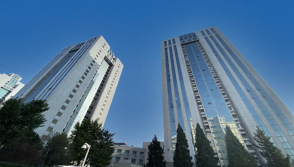
Beijing Saldi Technology Co., Ltd. is located in Shangdi Information Industry Base, Haidian District, Beijing. Since its establishment in 2009, the company has been committed to the introduction, consultation and distribution of scientific imaging products, and provides customers with scientific imaging solutions.
Dozens of industry applications have helped more than 1000 enterprises achieve stable standards.
In order to provide professional, advanced, efficient, innovative, and economically diverse solutions to our customers, the company acts as an agent to sell major scientific imaging industry brand products in Europe, America, Asia, and other regions, mastering advanced industry standards and technical experience. The products represented by our company have all passed the EU CE quality certification and are exported to major research institutes, universities, and technology enterprises both domestically and internationally, receiving the favor of our customers.
010-82895362
Website:http://meiz.hqceshi.vip/
Email:imaging@scientificimaging.cn
Address:Rm1-1705, Guoji Keji Chuangye Garden, No.1 Shangdi Xinxi Road, Haidian District, Beijing PRC


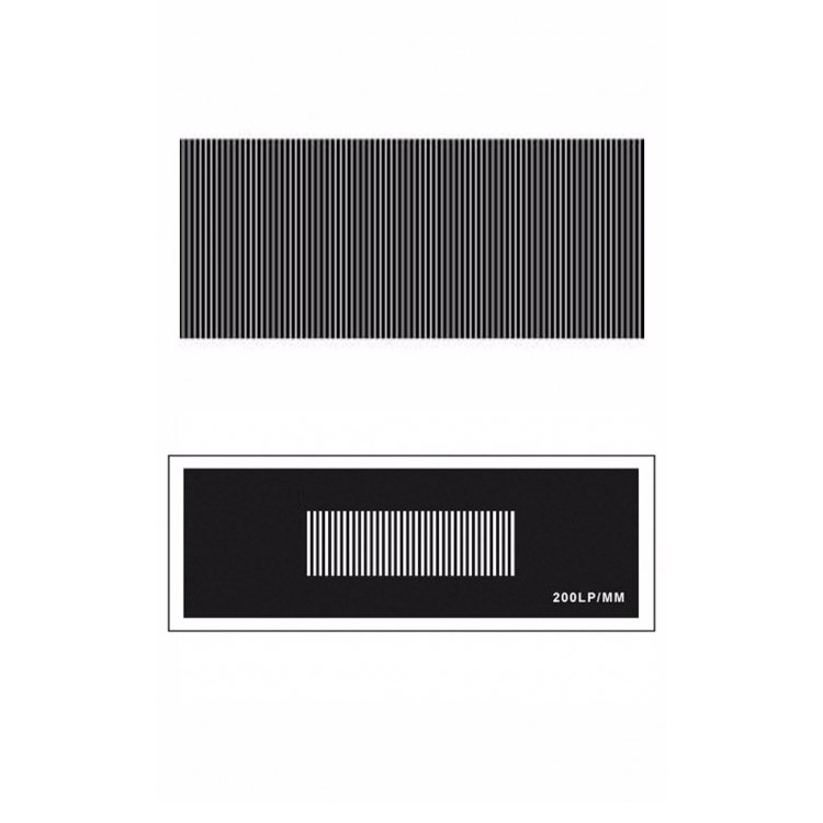


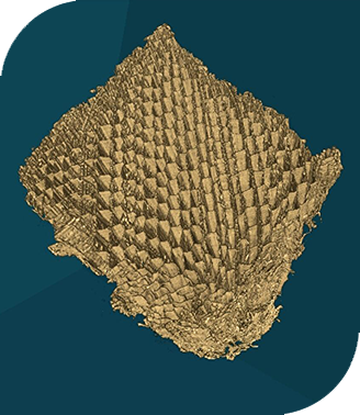
 X-ray imaging
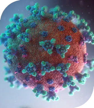
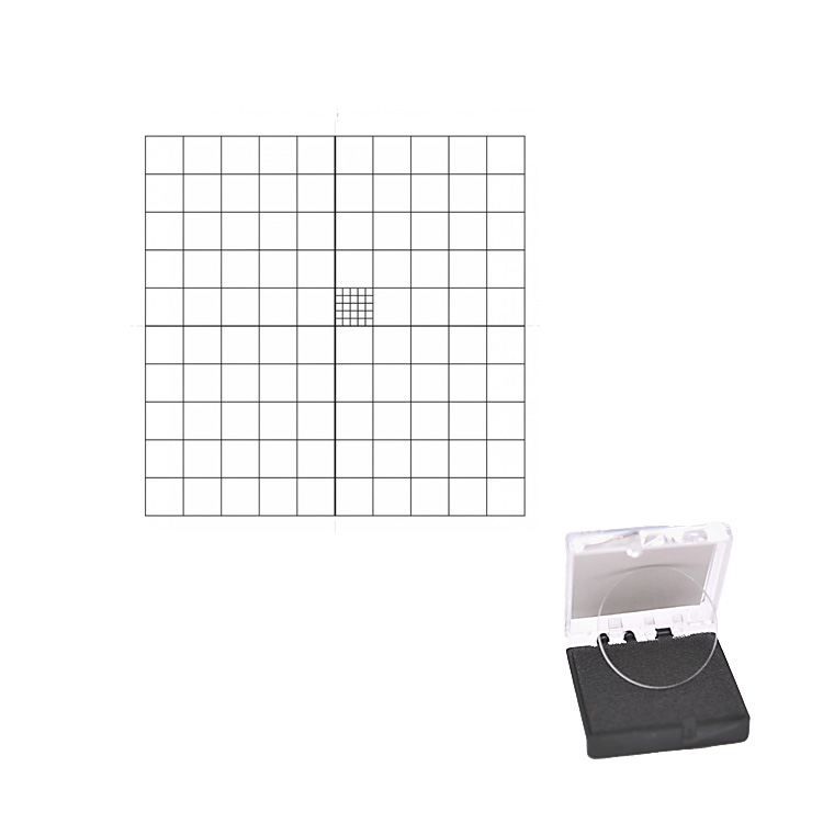
 Life sciences
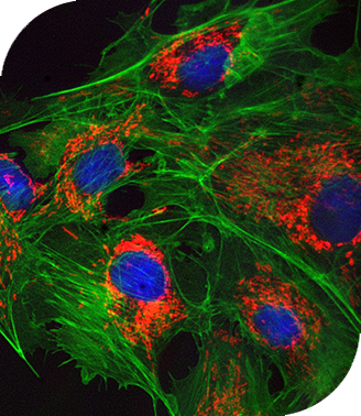
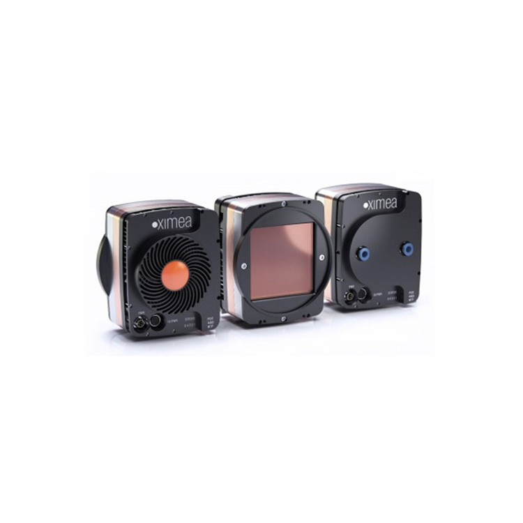
 Fluorescence imaging
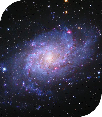
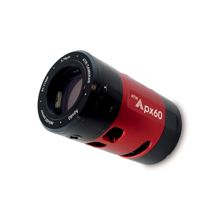
 Astronomical photography